Scientists reveal why some brains stop growing too soon
A single mutation in a cell’s internal scaffolding can derail early brain development.
- Date:
- December 17, 2025
- Source:
- Deutsches Primatenzentrum (DPZ)/German Primate Center
- Summary:
- Researchers used miniature human brains grown in the lab to uncover why certain genetic mutations lead to abnormally small brains. Changes in actin disrupted the orientation of early brain cell divisions, causing crucial progenitor cells to disappear too soon. This reduced the brain’s ability to grow normally. The work offers a clear cellular explanation for microcephaly linked to Baraitser-Winter syndrome.
- Share:
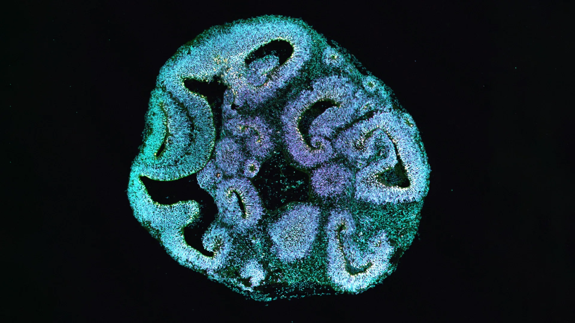
Why do some children develop a brain that is unusually small (microcephaly)? A global team of scientists from the German Primate Center -- Leibniz Institute for Primate Research (DPZ), Hannover Medical School (MHH), and the Max Planck Institute of Molecular Cell Biology and Genetics set out to answer this question using human brain organoids. These lab-grown models allowed the researchers to closely examine how changes in key structural proteins inside cells can interfere with early brain development.
Their work, documented in EMBO Reports, shows that mutations in actin genes disrupt how early brain progenitor cells divide. When these cells fail to divide correctly, their numbers drop, limiting overall brain growth and resulting in a smaller brain. "Our findings provide the first cellular explanation for microcephaly in people with the rare Baraitser-Winter syndrome," says Indra Niehaus, first author of the study and research associate at Hannover Medical School.
How the Cell's Internal Framework Shapes Brain Development
Actin plays a central role in the cytoskeleton, the internal framework that gives cells structure and helps move materials inside them. In people with Baraitser-Winter syndrome, a mutation affects one of two crucial actin genes. To understand the consequences, the researchers reprogrammed skin cells from affected patients into induced pluripotent stem cells. These stem cells were then used to grow three-dimensional brain organoids that mimic early stages of human brain formation.
After thirty days of development, the differences were striking. Organoids grown from patient cells were about 25 percent smaller than those grown from healthy donor cells. The ventricle-like regions inside the organoids, where progenitor cells gather and begin forming early nerve cells, were also much smaller.
A Shift in Crucial Brain Cell Populations
When the scientists examined the types of cells inside the organoids, they found a clear imbalance. The number of apical progenitor cells, which are essential for building the cerebral cortex, was significantly lower. At the same time, there was an increase in basal progenitor cells, which usually appear later as development progresses.
This shift suggested that the timing and outcome of cell division had been altered, potentially explaining why the brain tissue failed to expand normally.
When Cell Division Orientation Goes Wrong
Using high-resolution microscopy, the team closely tracked how apical progenitor cells divided. Under normal conditions, these cells divide mainly at right angles to the ventricular surface. This orientation ensures that cellular components are evenly shared and that two new apical progenitor cells are produced.
In organoids carrying the actin mutation, this pattern changed dramatically. Vertical divisions became far less common, while horizontal and angled divisions dominated. As a result, apical progenitor cells were less able to renew themselves. They detached from the ventricular zone more often and were more likely to become basal progenitor cells instead.
"Our analyses show very clearly that a change in the division orientation of the progenitor cells is the decisive trigger for the reduced brain size," says Michael Heide, group leader at the German Primate Center and last author of the study. "A single change in the cytoskeleton is sufficient to disrupt the course of early brain development."
Tiny Structural Changes With Lasting Effects
Electron microscopy revealed additional, subtle defects at the ventricular surface. Cell shapes appeared uneven, and extra protrusions formed between neighboring cells. Researchers also observed unusually high levels of tubulin at cell junctions. Tubulin is another cytoskeletal protein that plays a key role in cell division.
Although the overall structure of the cells remained intact, these small abnormalities may be enough to permanently alter how cells orient themselves during division.
Proving the Mutation Is the Cause
To confirm that the observed differences were truly caused by the actin mutation and not by other genetic variations, the researchers performed a crucial control experiment. They used CRISPR/Cas9 to introduce the exact same mutation into a healthy stem cell line. Brain organoids grown from these edited cells developed the same defects seen in patient-derived organoids -- a proof that the mutation itself is the driving factor.
What This Discovery Means for Medicine
The findings shed light on how rare genetic mutations can lead to complex brain malformations and demonstrate the value of brain organoids in biomedical research. "Our findings help us understand how rare genetic disorders lead to complex brain malformations and highlight the potential of brain organoids for biomedical research," says Michael Heide.
"The therapeutic potential of this study lies in diagnostics, as our data helps to better classify genetic findings in patients. Since the disease affects early fetal development processes, interventions in humans would be complex. However, new drugs that influence the interaction between actin and microtubules could open up new approaches in the long term," says Nataliya Di Donato, Director of the Institute of Human Genetics at Hannover Medical School.
Story Source:
Materials provided by Deutsches Primatenzentrum (DPZ)/German Primate Center. Note: Content may be edited for style and length.
Journal Reference:
- Indra Niehaus, Michaela Wilsch-Bräuninger, Felipe Mora-Bermúdez, Fabian Rost, Mihaela Bobic-Rasonja, Velena Radosevic, Marija Milkovic-Perisa, Pauline Wimberger, Mariasavina Severino, Alexandra Haase, Ulrich Martin, Karolina Kuenzel, Kaomei Guan, Katrin Neumann, Noreen Walker, Evelin Schröck, Natasa Jovanov-Milosevic, Wieland B Huttner, Nataliya Di Donato, Michael Heide. Cerebral organoids expressing mutant actin genes reveal cellular mechanism underlying microcephaly. EMBO Reports, 2025; DOI: 10.1038/s44319-025-00647-7
Cite This Page: