Scientists capture flu viruses surfing into human cells in real time
- Date:
- December 4, 2025
- Source:
- ETH Zurich
- Summary:
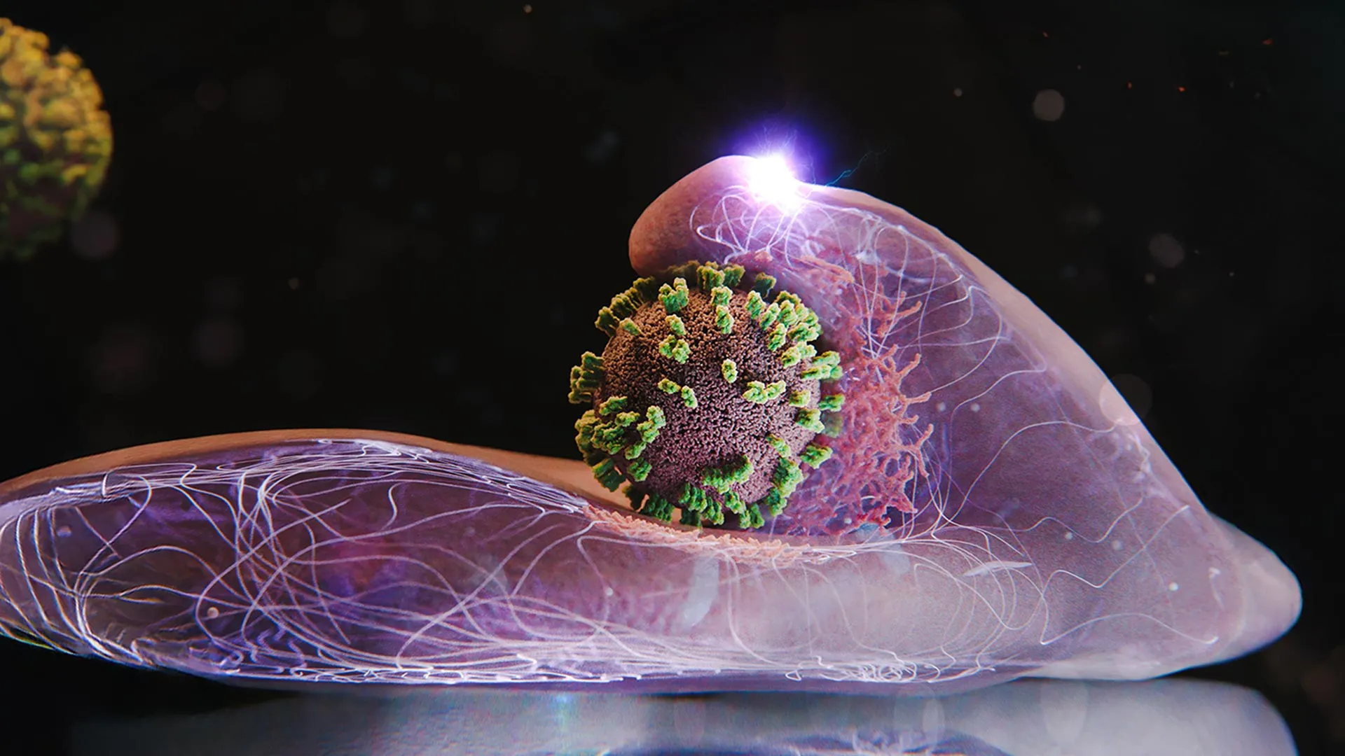
- Scientists have captured a never-before-seen, high-resolution look at influenza’s stealthy invasion of human cells, revealing that the cells aren’t just helpless victims. Using a groundbreaking imaging technique, researchers discovered that our cells actually reach out and “grab” the virus as it searches for the perfect entry point, surfing along the membrane.
- Share:
Fever, aching limbs and a runny nose -- as winter returns, so too does the flu. The disease is triggered by influenza viruses, which enter our body through droplets and then infect vulnerable cells.
A research team from Switzerland and Japan has taken an exceptionally close look at how this virus behaves. Using a microscopy approach they created themselves, the scientists can zoom in on the outer surface of human cells in a Petri dish. This setup has enabled them to watch, live and in sharp detail, the moment an influenza virus penetrates a living cell.
Under the direction of Yohei Yamauchi, Professor of Molecular Medicine at ETH Zurich, the group discovered something unexpected. The cells do not simply sit idle while the influenza virus approaches. Instead, they appear to make an effort to seize it. "The infection of our body cells is like a dance between virus and cell," says Yamauchi.
Viral Surfing on the Cell Surface
Although cells gain nothing from being infected, the interaction looks active because the virus exploits a routine cellular uptake system that the cells cannot do without. This system normally brings essential substances such as hormones, cholesterol or iron into the cell.
To begin infection, an influenza virus attaches to specific molecules on the cell surface. The process resembles surfing on the membrane. The virus moves along the surface, latching onto one molecule after another, until it arrives at a site rich in these receptors. A spot with many receptors side by side provides the most efficient entry route.
When the cell's receptors detect that the virus has attached, the membrane begins forming a small indentation at that spot. A structural protein named clathrin shapes and supports this deepening pocket. As the pocket expands, it wraps around the virus and forms a vesicle. The cell then pulls this vesicle inward, where the coat dissolves and releases the virus.
Why Earlier Microscopy Fell Short
Previous attempts to study this crucial moment in infection relied on methods like electron microscopy, which require destroying the cells to obtain an image. As a result, they captured only single moments in time. Fluorescence microscopy, another common tool, offers live imaging but at low spatial resolution.
ViViD-AFM Sheds Light on Viral Entry
The new method, which merges atomic force microscopy (AFM) with fluorescence microscopy, is called virus-view dual confocal and AFM (ViViD-AFM). This combined approach makes it possible to track the fine-scale movements involved as the virus enters the cell.
With this tool, the researchers demonstrated that cells assist the virus at several stages of entry. They summon important clathrin proteins to the site where the virus is attached. The membrane at that point also pushes upward, almost as if trying to seize the virus. These wave-like motions intensify if the virus tries to drift away from the surface.
Implications for Antiviral Research
Because ViViD-AFM allows scientists to observe infection while it is happening, it offers a valuable way to test antiviral drug candidates directly in cell cultures. The team notes that the technique may also be applied to studying other viruses or even vaccines, giving researchers a real-time view of how these particles interact with cells.
Story Source:
Materials provided by ETH Zurich. Note: Content may be edited for style and length.
Journal Reference:
- Aiko Yoshida, Yoshitsugu Uekusa, Takeshi Suzuki, Michael Bauer, Nobuaki Sakai, Yohei Yamauchi. Enhanced visualization of influenza A virus entry into living cells using virus-view atomic force microscopy. Proceedings of the National Academy of Sciences, 2025; 122 (38) DOI: 10.1073/pnas.2500660122
Cite This Page: