
MIT scientists build a tiny robot that swims using living muscle cells
- Date:
- October 23, 2026
- Source:
- Massachusetts Institute of Technology
- Summary:
- MIT engineers have built a paper-thin swimming robot powered by living muscle cells that respond to flashes of light, allowing it to swim and steer through water. The breakthrough could pave the way for tiny, self-healing biological robots capable of exploring fragile environments that ordinary machines cannot safely navigate.
- Share:
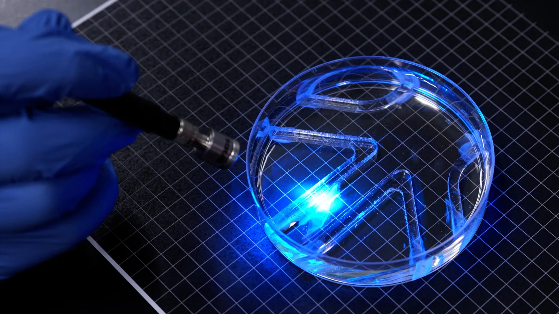
A swimming robot does not necessarily need a bulky motor or powerful mechanical parts to move through water. MIT researchers have demonstrated that a single, carefully engineered layer of living muscle cells can generate enough force to propel a tiny robot forward.
The engineers developed an extremely thin swimming robot that uses living muscle tissue to move its fins. Described in a new study published in Advanced Functional Materials, the device has a flexible gel framework roughly the length and width of a stick of gum. The gel forms two fins, each coated with a layer of muscle cells far thinner than a human hair. These cells have been genetically modified to contract when exposed to light.
By directing light onto either fin, the researchers can trigger muscle contractions that make the fin flap and push the robot through water. Adjusting which fin receives light and how frequently it is illuminated allows the team to control both its speed and direction.
In experiments, the remarkably thin robot successfully swam and turned through a simple underwater maze. At maximum speed, it covered approximately four times its body length per minute. Although that is much slower than an Olympic swimmer, who can travel up to 65 body lengths per minute, it is comparable to the swimming pace of a cow shark moving leisurely through the ocean.
"It takes a lot of force to move through water versus air," says study author Ritu Raman, associate professor of mechanical engineering at MIT. "The robot's quite strong, given its size. "
A New Generation of Robots Powered by Living Muscle
The device represents the first demonstration of a very thin, two-dimensional robot that can move from place to place using living muscle tissue.
Most existing biohybrid robots rely on relatively large, three-dimensional structures made from laboratory-grown skeletal muscle. Producing these structures typically requires millions of cells. Raman explains that thinner designs could reduce manufacturing costs while potentially improving movement efficiency.
"Currently, biohybrid robots from our group and others' are built from bulky, 3D chunks of lab-grown skeletal muscle that require millions of cells to fabricate," says Raman, who notes that thinner, less bulky designs such as the team's new bot could be cheaper to build and could move more efficiently. "We believe that biohybrid robots powered by living muscle could one day perform delicate jobs like exploring environments too fragile or unpredictable for conventional hardware, because living tissue is soft, responsive to its surroundings, and can heal itself."
The MIT research team also included first author Maheera Bawa, along with Arielle Berman, Laura Schwendeman, Ferdows Afghah, and Seanbiron Johnson.
From Artificial Muscle to a Swimming Robot
The new swimming robot builds on an earlier achievement by Raman's laboratory. Last year, the researchers created an artificial muscle disk inspired by the human iris, demonstrating how a thin layer of living cells could produce precisely controlled movements.
For that project, the engineers patterned a gel disk with grooves extending outward from its center and forming concentric circles. They then placed living muscle cells on the surface, where the cells grew into a thin layer following the patterned channels.
When illuminated, the muscle tissue contracted in different directions, causing the disk to stretch and compress. Its movements resembled the way the iris of the human eye changes shape as the pupil expands or narrows.
The experiment established that researchers could grow extremely thin muscle layers in intricate arrangements and use light to control their movements along multiple directions.
"People hadn't seen this muscle architecture engineered from scratch before," Raman says. "And the cells were moving in multiple directions. But they only moved about 100 microns. From a robotics perspective, their movements were tiny."
For the latest project, the researchers wanted to dramatically increase the amount of movement and force generated by the muscle cells. Their goal was to produce contractions powerful enough to propel a robot through water.
They determined that achieving this would require a better foundation for the living muscle tissue.
The earlier artificial iris had used fibrin, an exceptionally soft gel, as its underlying structure. However, the team found that fibrin could quickly shrink under the forces exerted by contracting muscle cells, limiting the effectiveness of the resulting movement.
To overcome this problem, the engineers investigated three characteristics of the gel foundation: its chemical composition, its stiffness, and the dimensions and geometry of the grooves used to organize muscle growth.
"For engineering any type of tissue, it's known that these are knobs you can tune," Raman says. "And we wanted to optimize all these parameters to support live muscle cells."
Engineering a Stronger Muscle-Powered Robot
Finding the right supporting material required testing gels with different formulations, stiffness levels, and surface patterns.
The researchers compared several groove shapes, including narrow channels with square profiles and longer channels with curved bottoms. They discovered that muscle cells aligned more effectively inside square-shaped grooves.
This alignment was important because muscle cells that grow in the same direction are more likely to merge into fibers. Those fibers can then form stronger muscle tissue that contracts in a coordinated way. The square grooves therefore provided a more effective structure for producing force.
The team also replaced fibrin with gelatin methacrylate (GelMA), a material commonly used to grow and engineer biological tissue.
By adjusting the formulation of GelMA, the researchers produced supporting gels with varying degrees of stiffness. They then examined how muscle cells developed on each surface and measured their ability to generate force.
Stiffer gels produced the best results. On these firmer surfaces, muscle cells grew in better alignment and generated stronger contractions.
The engineers next investigated how thick the gel should be. They determined that a film measuring approximately half a millimeter offered an effective balance. It was sturdy enough to support a single layer of muscle cells while remaining sufficiently lightweight to prevent the contracting cells from detaching from the surface.
The team further improved muscle performance through a form of exercise. Repeated pulses of light stimulated the cells to contract, strengthening the muscle tissue before it was incorporated into the swimming robot.
How Light Makes the Living Robot Swim
With the gel structure optimized and the muscle cells strengthened, the researchers assembled their swimming device.
The finished robot consisted of a thin gel framework divided into two fins. Both sides of the gel were patterned with square-bottomed grooves and covered with living muscle cells. As the cells developed, they fused into organized muscle fibers capable of contracting together.
Each fin could be activated independently using light, giving the researchers a simple way to control the robot's movements.
"You can think of the robot as having two independent muscles," Raman says. "If we shine a light on just one, only that muscle moves. If shining on both, they both flap."
To test the device, the researchers placed it inside a large petri dish filled with water. They then moved a light source by hand, illuminating the fins to guide the robot through a maze positioned inside the dish.
As the muscles contracted, the fins flapped and propelled the robot through the water. By changing the light stimulation, the engineers successfully controlled its movement and helped it navigate the obstacles.
Although the robot demonstrated the ability to swim and steer, its current shape remains relatively simple. The primary objective of the experiment was to establish that an exceptionally thin layer of engineered muscle tissue could generate enough force for swimming.
The researchers now plan to refine the robot's body to improve its speed. Beyond demonstrating a new approach to biohybrid robotics, the technology could eventually be useful for monitoring aquatic environments or operating in locations where conventional rigid machines might cause damage.
"Our next goal is to optimize the body design to enable faster swimming," Raman says. "But even at slow swim speeds, one could imagine a muscle-powered swimmer being used for purposes like environmental monitoring in aquatic environments."
The Office of Naval Research provided partial funding for this research.
Story Source:
Materials provided by Massachusetts Institute of Technology. Original written by Jennifer Chu. Note: Content may be edited for style and length.
Journal Reference:
- Maheera Bawa, Arielle Berman, Laura Schwendeman, Ferdows Afghah, Seanbiron Johnson, Ritu Raman. 2D Skeletal Muscle Thin Film Actuators Enhance Efficiency of Biohybrid Robots. Advanced Functional Materials, 2026; DOI: 10.1002/adfm.78065
Cite This Page: