Breakthrough microchip reveals how your body fights viruses—in just 90 minutes
By analyzing just a drop of blood, this microchip gives researchers quicker-than-ever insight into how a person’s antibodies are interacting with a virus or other pathogen.
- Date:
- July 12, 2025
- Source:
- Scripps Research Institute
- Summary:
- A team at Scripps Research has created a microchip that can rapidly reveal how a person's antibodies respond to viruses using only a drop of blood. This game-changing technology, called mEM, condenses a week’s worth of lab work into 90 minutes, offering a powerful tool for tracking immune responses and fast-tracking vaccine development. Unlike earlier methods, it needs far less blood and delivers more detailed insights, even revealing previously undetected antibody targets on viruses like SARS-CoV-2 and influenza.
- Share:
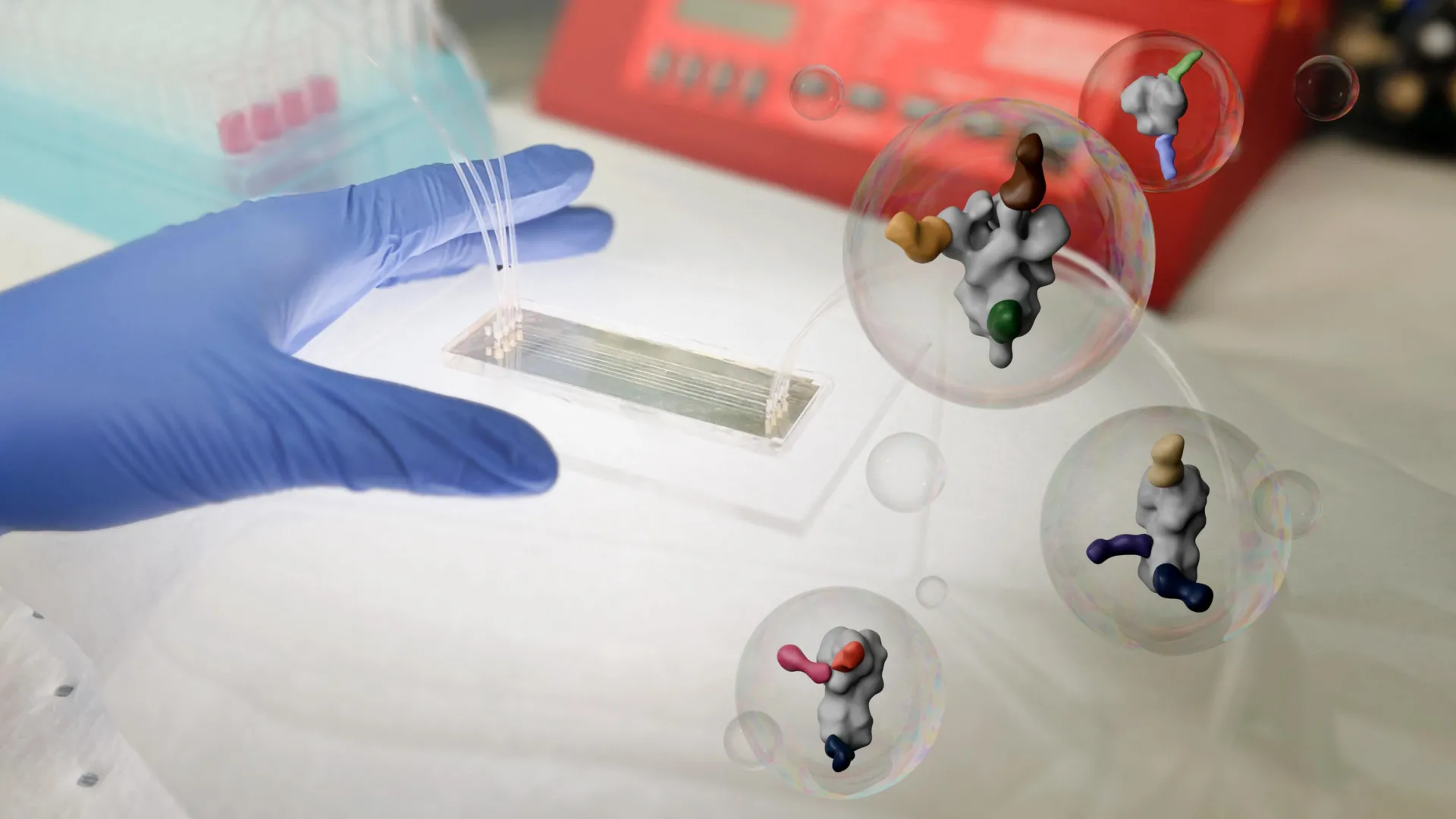
A new microchip invented by Scripps Research scientists can reveal how a person's antibodies interact with viruses -- using just a drop of blood. The technology offers researchers faster, clearer insights that could help accelerate vaccine development and antibody discovery.
"This lets us take a quick snapshot of antibodies as they are evolving after a vaccine or pathogen exposure," says Andrew Ward, professor in the Department of Integrative Structural and Computational Biology at Scripps Research and senior author of the new paper published in Nature Biomedical Engineeringon June 3, 2025. "We've never been able to do that on this timescale or with such tiny amounts of blood before."
When someone is infected with a virus, or receives a vaccine, their immune system creates new antibodies to recognize the foreign invader. Some antibodies work well against the pathogen, while others attach to it only weakly. Figuring out exactly which parts of the virus the best antibodies stick to is key information for scientists trying to optimize vaccines, since they want to design vaccines that elicit strong, reliable immune responses.
"If we know which particular antibodies are leading to the most protective response against a virus, then we can go and engineer new vaccines that elicit those antibodies," says Leigh Sewall, a graduate student at Scripps Research and first author of the new paper.
In 2018, Ward's lab unveiled a technique known as electron microscopy-based polyclonal epitope mapping (EMPEM). This method allowed scientists to visualize how antibodies in blood samples attach to a virus. Although groundbreaking, it had downsides: it took a full week to complete and required relatively large amounts of blood.
"During the COVID-19 pandemic, we began really wanting a way to do this faster," says Alba Torrents de la Peña, a Scripps Research staff scientist who helped lead the work. "We decided to design something from scratch."
With the new system, known as microfluidic EM-based polyclonal epitope mapping (mEM), researchers start with four microliters of blood extracted from a human or animal-about one hundred times less than what's required in original EMPEM. The blood is injected in a tiny, reusable chip where viral proteins are stuck to a special surface. As the blood flow through the chip, antibodies recognize and bind to those. Then, the viral proteins -- with any antibodies attached -- are gently released from the chip and prepared for imaging using standard electron microscopy. The entire process only takes about 90 minutes.
To test the value and effectiveness of mEM, the research team used the system to map antibodies in humans and mice that had either received a vaccination against or been infected with a virus, including influenza, SARS-CoV-2 and HIV. The new technique was not only fast at mapping out the interactions between antibodies and those viruses, but more sensitive than EMPEM; it revealed new antibody binding sites on both influenza and coronavirus proteins that had not been picked up by EMPEM.
To track how antibodies evolved over time in individual mice after they received a vaccination against one of the pathogens, the team took small blood samples from a mouse at different time points.
"That was something that wouldn't have been possible in the past, because of the amount of blood needed for EMPEM," says Sewall. "So to be able to look at an individual over time was really exciting."
The researchers are now working to automate and multiplex the system, which could eventually allow dozens of samples to be processed in parallel. Ultimately, they envision mEM becoming a widely adopted tool to monitor and guide vaccine development in pathogens ranging from coronaviruses to malaria.
"This technology is useful in any situation where you have really limited sample volume, or need initial results quickly," says Torrents de la Peña. "We hope this becomes accessible to more researchers as it is simplified and streamlined."
In addition to Ward, Sewall, and Torrents de la Peña, authors of the study, "Microfluidics Combined with Electron Microscopy for Rapid and High-Throughput Mapping of Antibody-Viral Glycoprotein Complexes," are Rebeca de Paiva Froes Rocha, Grace Gibson, Michelle Louie, Sandhya Bangaru, Andy S. Tran, Gabriel Ozorowski, Blanca Chocarro Ruiz, Nathan Beutler, Thomas F. Rogers, Dennis R. Burton, and Andrew B. Ward of The Scripps Research Institute; Zhenfei Xie and Facundo D. Batista of the Ragon Institute of MGH, MIT and Harvard; and Subhasis Mohanty and Albert C. Shaw of Yale University School of Medicine.
This work was supported by funding from the National Institutes of Health (AI136621, AI089992, and AI144462), and by the Bill and Melinda Gates Foundation (INV-002916).
Story Source:
Materials provided by Scripps Research Institute. Note: Content may be edited for style and length.
Journal Reference:
- Leigh M. Sewall, Rebeca de Paiva Froes Rocha, Grace Gibson, Michelle Louie, Zhenfei Xie, Sandhya Bangaru, Andy S. Tran, Gabriel Ozorowski, Subhasis Mohanty, Nathan Beutler, Thomas F. Rogers, Dennis R. Burton, Albert C. Shaw, Facundo D. Batista, Blanca Chocarro Ruiz, Alba Torrents de la Peña, Andrew B. Ward. Microfluidics combined with electron microscopy for rapid and high-throughput mapping of antibody–viral glycoprotein complexes. Nature Biomedical Engineering, 2025; DOI: 10.1038/s41551-025-01411-x
Cite This Page: