MIT scientists finally reveal the hidden structure of a mysterious high-tech material
A long-standing materials mystery is solved—unlocking a path to smarter, more powerful tech.
- Date:
- May 4, 2026
- Source:
- Massachusetts Institute of Technology
- Summary:
- For decades, relaxor ferroelectrics have powered everything from medical ultrasounds to sonar systems, yet their inner atomic structure remained a mystery—until now. Researchers have finally mapped their three-dimensional structure in unprecedented detail, uncovering hidden patterns in how electric charges are arranged at the nanoscale. The breakthrough not only challenges long-standing assumptions about how these materials behave but also allows scientists to refine the models used to design them.
- Share:
Materials known as relaxor ferroelectrics have played an important role for decades in technologies such as ultrasound imaging, microphones, and sonar. Their unusual performance comes from the way atoms are arranged inside them. However, that internal structure has been extremely difficult to measure directly, leaving scientists to rely on incomplete models.
Now, researchers from MIT and collaborating institutions have, for the first time, mapped the three dimensional atomic structure of a relaxor ferroelectric. Their results, to be published in Science, offer a clearer foundation for improving the models used to design future computing systems, energy devices, and advanced sensors.
"Now that we have a better understanding of exactly what's going on, we can better predict and engineer the properties we want materials to achieve," says corresponding author James LeBeau, MIT's Kyocera Professor of Materials Science and Engineering. "The research community is still developing methods to engineer these materials, but in order to predict the properties those materials will have, you have to know if your model is right."
Revealing Hidden Charge Patterns in Complex Materials
In the study, the team used a cutting edge imaging method to examine how electric charges are distributed throughout the material. What they found challenged previous assumptions.
"We realized the chemical disorder we observed in our experiments was not fully considered previously," says co-first authors Michael Xu PhD '25 and Menglin Zhu, who are both postdocs at MIT. "Working with our collaborators, we were able to merge the experimental observations with simulations to refine the models and better predict what we see in experiments."
The research team also included Colin Gilgenbach and Bridget R. Denzer, MIT PhD students in materials science and engineering; Yubo Qi, an assistant professor at the University of Alabama at Birmingham; Jieun Kim, an assistant professor at the Korea Advanced Institute of Science and Technology; Jiahao Zhang, a former PhD student at the University of Pennsylvania; Lane W. Martin, a professor at Rice University; and Andrew M. Rappe, a professor at the University of Pennsylvania.
Probing Disordered Materials at the Atomic Scale
Computer models have long suggested that when an electric field is applied to relaxor ferroelectrics, interactions between positively and negatively charged atoms within tiny regions help create their strong energy storage and sensing abilities. Until now, those nanoscale regions could not be directly observed.
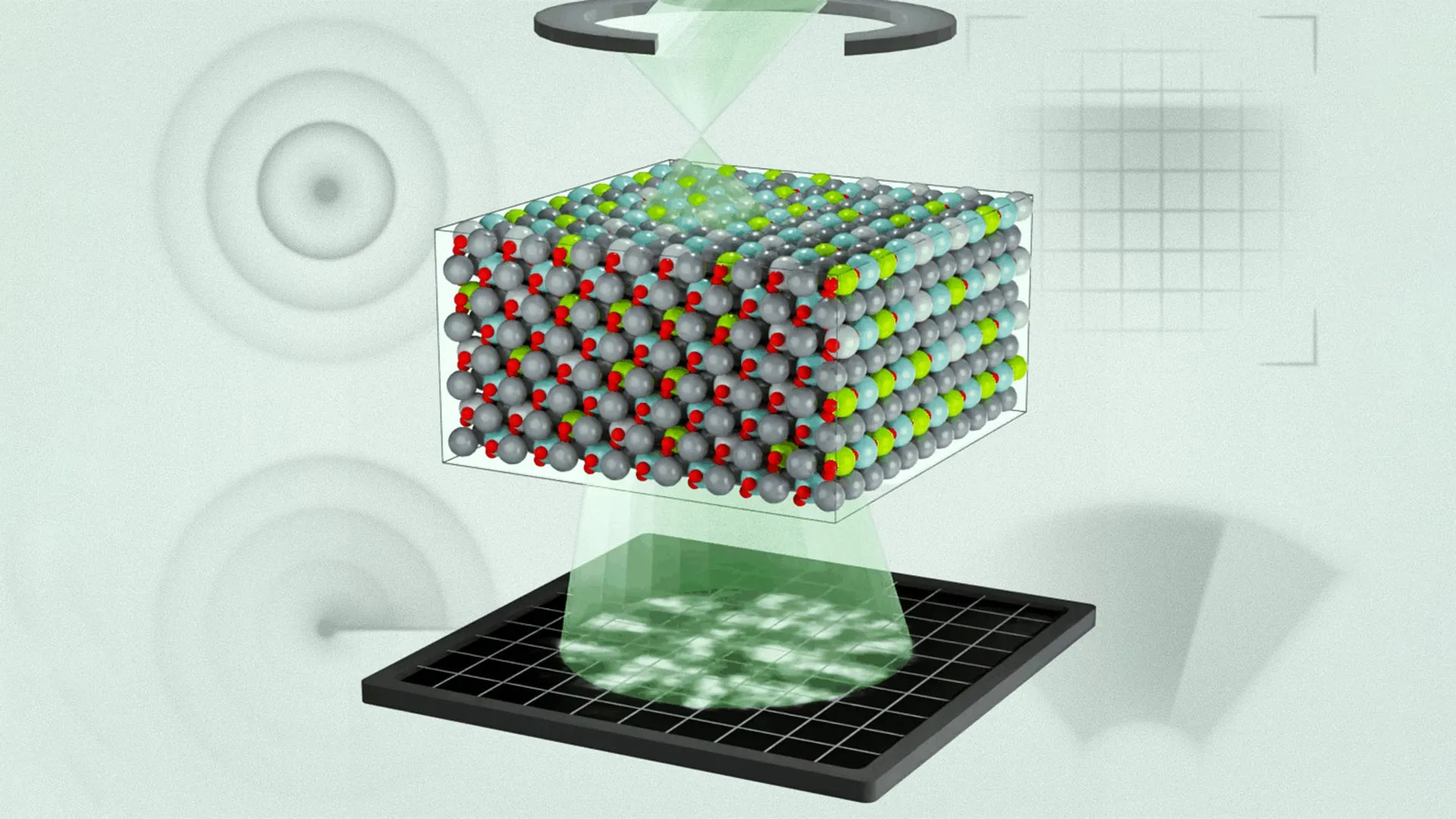
To investigate further, the researchers focused on a widely used material found in sensors, actuators, and defense systems, a lead magnesium niobate-lead titanate alloy. They applied an advanced technique called multi-slice electron ptychography (MEP). This method involves scanning a nanoscale beam of high energy electrons across the material and recording the diffraction patterns that result.
"We do this in a sequential way, and at each position, we acquire a diffraction pattern," Zhu explains. "That creates regions of overlap, and that overlap has enough information to use an algorithm to iteratively reconstruct three-dimensional information about the object and the electron wave function."
Using this approach, the team uncovered a layered hierarchy of chemical and polar structures, extending from individual atoms up to larger, mesoscopic features. They also discovered that regions with different polarization were significantly smaller than earlier simulations had predicted. By incorporating these observations into their models, the researchers were able to improve how well simulations match real world behavior.
"Previously, these models basically had random regions of polarization, but they didn't tell you how those regions correlate with each other," Xu says. "Now we can tell you that information, and we can see how individual chemical species modulate polarization depending on the charge state of atoms."
Toward Better Materials for Future Technologies
According to Zhu, the findings highlight the growing power of electron ptychography for exploring complex, disordered materials and could lead to new lines of research.
"This study is the first time in the electron microscope that we've been able to directly connect the three-dimensional polar structure of relaxor ferroelectrics with molecular dynamics calculations," Xu says. "It further proves you can get three-dimensional information out of the sample using this technique."
The team believes this method could eventually help scientists design materials with tailored electronic properties, improving technologies such as memory storage, sensing systems, and energy devices.
"Materials science is incorporating more complexity into the material design process -- whether that's for metal alloys or semiconductors -- as AI has improved and our computational tools have become more advanced," LeBeau says. "But if our models aren't accurate enough and we have no way to validate them, it's garbage in garbage out. This technique helps us understand why the material behaves the way it does and validate our models."
The research was supported in part by the U.S. Army Research Laboratory, the U.S. Office of Naval Research, the U.S. Department of War, and a National Science Graduate Fellowship. The work also made use of MIT.nano facilities.
Story Source:
Materials provided by Massachusetts Institute of Technology. Original written by Zach Winn. Note: Content may be edited for style and length.
Journal Reference:
- Menglin Zhu, Michael Xu, Yubo Qi, Colin Gilgenbach, Jieun Kim, Jiahao Zhang, Bridget R. Denzer, Lane W. Martin, Andrew M. Rappe, James M. LeBeau. Bridging experiment and theory of relaxor ferroelectrics with multislice electron ptychography. Science, 2026; 392 (6797): 519 DOI: 10.1126/science.ads6023
Cite This Page: